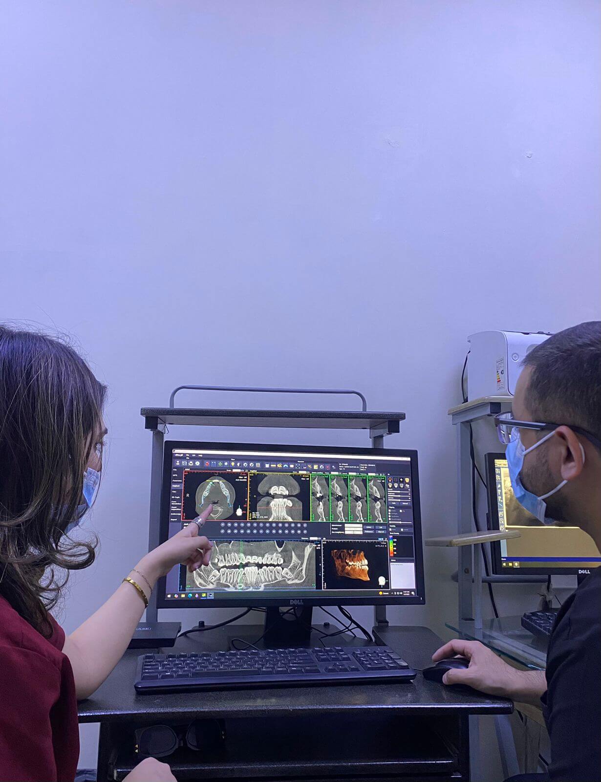
Dental implant surgery has come a long way. What used to involve a significant amount of estimation and clinical guesswork is now guided by imaging technology precise enough to show a dentist exactly what they’re working with before they’ve made a single incision.
CBCT (Cone Beam Computed Tomography) is the technology behind that shift. And in complex implant cases, it’s become less of a premium add-on and more of a clinical standard for dentists who take the outcome seriously.
Here’s why.
What CBCT Actually Is
CBCT is a specialized imaging system that produces three-dimensional scans of the jaw, teeth, bone, and surrounding structures. Unlike a conventional dental X-ray, which gives you a flat, two-dimensional picture, CBCT gives the dentist a complete volumetric view, every angle, every layer, every anatomical detail that matters for planning a procedure.
For dental implant surgery, that difference in information quality is significant. The more a dentist can see before the procedure begins, the more accurately they can plan, and the less they’re relying on assumptions that could affect the outcome.
1. It Shows Bone Volume and Density in Three Dimensions
The foundation of a successful implant is the bone it sits in. Too little bone volume or insufficient density, and the implant won’t integrate properly, leading to failure, the need for bone grafting, or both.
A conventional X-ray gives a two-dimensional estimate of bone height. CBCT gives a complete three-dimensional picture of bone volume, height, width, and density, the full information set a dentist needs to determine whether a patient can proceed directly to dental implant surgery, whether bone grafting is required first, and what implant size and positioning will give the best chance of long-term success.
In complex cases: multiple missing teeth, previous bone loss, patients with a history of periodontal disease, this level of detail isn’t optional. It’s the difference between a treatment plan built on complete information and one built on incomplete information.
2. It Identifies Critical Anatomical Structures
The jaw contains structures that a dentist absolutely cannot afford to disturb during dental implant surgery: the inferior alveolar nerve in the lower jaw, the maxillary sinus in the upper jaw, and blood vessels that run close to the implant site.
On a standard X-ray, the exact position of these structures relative to the proposed implant site involves a degree of estimation. On a CBCT scan, their precise three-dimensional location is visible, which means the dentist can plan the implant position, angle, and depth to maintain a safe margin from each structure.
For patients pursuing permanent tooth replacement in areas close to the nerve canal or sinus floor, this precision isn’t just clinically important; it’s what prevents complications that, in worst cases, can include nerve damage or sinus perforation.
3. It Enables Accurate Surgical Guide Fabrication
One of the most significant advances in implant dentistry in recent years is the surgical guide. This custom-made template fits over the patient’s teeth and directs the drill to exactly the right position and angle during surgery.
Surgical guides can only be as accurate as the imaging they’re built from. CBCT data, combined with digital planning software, allows a dentist to plan the entire implant procedure virtually before the patient sits in the chair, placing the implant on screen, checking every clearance, and then fabricating a guide that translates that plan precisely into the actual surgery.
For complex cases involving multiple implants or anatomically challenging sites, this level of pre-surgical planning makes the procedure faster, more predictable, and significantly safer.
4. It Supports Better Outcomes for Full Arch Permanent Tooth Replacement
Full arch permanent tooth replacement (replacing all teeth in the upper or lower jaw) is one of the most technically demanding procedures in implant dentistry. The number of implants involved, the distribution of bone across the arch, and the need for each implant to be positioned correctly for the final prosthesis to fit and function properly all require a level of planning that goes well beyond what a standard X-ray supports.
CBCT allows the dentist and the laboratory to plan the full arch case as a complete system, understanding how bone volume and distribution will affect implant positions, how those positions will support the final prosthetic design, and where any challenges need to be addressed before surgery begins.
The patients who benefit most from this level of planning are exactly the patients most likely to need full arch replacement: those who have lost teeth over a long period, often with accompanying bone loss, who need a solution that works within the anatomical constraints of what remains.
5. It Reduces Surprises During Surgery
In surgery, surprises are rarely good. An unexpected anatomical variation, a root that wasn’t visible on a conventional X-ray, a bone density that differs significantly from what was estimated, each of these can change what should be a straightforward procedure into a more complicated one.
CBCT virtually eliminates this category of surprise. What can be seen has been seen, in three dimensions, before the procedure begins. The dentist walks into surgery with a complete picture rather than a partial one, which means decisions can be made in the planning stage, where there’s time and information to make them well, rather than intraoperatively.
At Altamash Dental Clinic, CBCT imaging is integrated into the planning process for complex dental implant surgery cases precisely because the information it provides changes what’s possible in terms of accuracy, safety, and predictable outcomes.
Conclusion
CBCT isn’t a technology that dentists use because it’s impressive. They use it because it makes their work more accurate and their patients’ outcomes more predictable. In complex implant cases, where bone is limited, anatomy is challenging, or multiple implants are involved, the information a CBCT scan provides is genuinely irreplaceable.
For patients considering permanent tooth replacement through dental implants, understanding that your dentist is using this level of imaging to plan your case is a reasonable thing to ask about. It’s a meaningful indicator of how seriously the procedure is being approached.
Visit Altamash Dental Clinic to discuss your implant case with a team that uses the technology and the planning process that complex cases actually require.
FAQs
Is CBCT scanning safe?
CBCT uses a significantly lower radiation dose than a medical CT scan. The dose is higher than a conventional dental X-ray, but the clinical benefit in complex implant cases and the precision of planning it enables justifies its use.
Does every implant case require a CBCT scan?
Not necessarily. CBCT becomes particularly important in complex cases: multiple implants, limited bone, proximity to critical structures, or full arch permanent tooth replacement.
How long does a CBCT scan take?
The scan itself takes less than a minute. Processing and analysis of the data takes longer, but the result is a complete three-dimensional dataset that the dentist can work with in planning software before recommending a treatment plan.